Preclinical Imaging Center (PIC)
The preclinical imaging center (PIC) directed by external page Prof. Daniel Razansky consolidates several research groups devoted to development of novel functional and molecular imaging methods. PIC possesses state-of-the-art infrastructure for in vivo imaging in mice and rats, including an independent animal housing facility located within the same hygiene area. Our multi-modality imaging capacities include advanced functional magnetic resonance (fMRI), multi-spectral optoacoustic tomography (MSOT) scanners, fluorescence and multi-photon microscopes, functional ultrasound and more. Some of the instruments and core methodologies were invented and built in our center thus have no analogues worldwide. The facilities are fully equipped with advanced animal surgical tools, anesthesia systems, physiology monitoring suites, high-end computational resources, and other key equipment for small animal imaging research. Our partners primarily originate from the biomedical research community within USZ, UZH and ETH Zürich and, to some extent, from academic and research institutions outside Zürich as well as industrial partners.
As part of its research framework, PIC provides access to high end 7T and 9.4T small-animal MRI scanners equipped with cryocoils. For more information and project template, please refer to our Download guidelines for initiation of collaborative projects with PIC.
Bruker Biospec 94/30

9.4 T, 30 cm bore size, RF shielded room, 20 cm whole body gradient insert, 11.8 cm head gradient insert, max. gradient 700 mT/m, rise time 77 µs, Avance III HD electronics, Paravision 6 software platform, Quad. T/R Cryocoil (mouse), 2x2 receive array Cryocoil (mouse), Several head, body and heart RF coils for mice and rats.
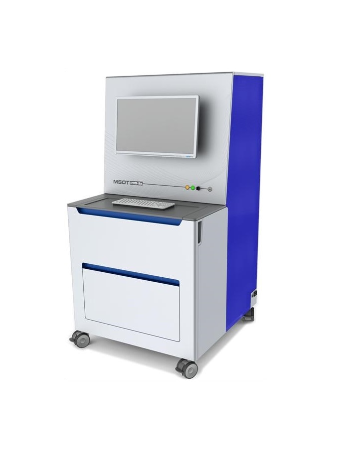
Multi-Spectral Optoacoustic Tomography (MSOT)

A number of commercial and custom-build preclinical multi-spectral optoacoustic tomography and scanning microscopy systems are available. Those are equipped with 2D and 3D ultrafast imaging capabilities (up to 100-1000 volumes per second), broad wavelength tunability between 420-1900nm and multi-scale imaging capacities from single cells to whole-organ and -body levels.
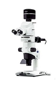
Fluorescence microscopy and macroscopy

Variety of high-end intravital multicolor fluorescence imaging systems are available, including multi-photon, wide-field, structured illumination, and optical coherence microscopes as well as macroscopic diffuse fluorescence tomography. The spectral coverage includes both visible and near-infrared (I and II) windows. The broad selection of systems allows for comprehensive in vivo investigations into multi-scale biological dynamics, such as brain-wide neural activity, vascularity and blood flow, molecular probe uptake, specific protein expression, circulation of cells or particles.
Contact
Inst. f. Biomedizinische Technik
Wolfgang-Pauli-Str. 27
8093
Zürich
Switzerland
